Converting CO2 into a valuable energy carrier using a model In2O3 catalyst

Converting CO2 into a valuable energy carrier using a model In2O3 catalyst

New discoveries made possible by In Situ TEM gas and heating

Original article by Athanasia Tsoukalou, Paula M. Abdala, Dragos Stoian, Xing Huang, Marc-Georg Willinger, Alexey Fedorov and Christoph R. Müller. Published in the Journal of the American Chemical Society on 19 July 2019.

Artist impression of methanol synthesis via CO2 hydrogenation using In2O3 catalyst (copyright of the American Chemical Society)

The direct hydrogenation of CO2 to methanol shows promise to be an important technique to reduce the amount of greenhouse gases in the atmosphere and thereby mitigate the negative effects of climate change while producing an important energy carrier. In his contribution to this article, Dr. Xing Huang has used In Situ TEM techniques to assess the limits of In2O3 catalytic performance in CO2 hydrogenation.

In Situ TEM Climate Nano-Reactor study

This catalyst research article by ETH Zürich provides a good demonstration on how in situ TEM experiments can add value to the study of reaction mechanisms. First, the structural changes in the catalyst material were studied with traditional TEM techniques before and after the CO2 reduction. By using the Climate in situ TEM system, the researchers were able to see the dynamics of the reaction, even at much lower pressures (0.8 vs. 20 Bar) than in the original operando experiment.

In Situ TEM video made using the DENSsolutions Climate system, at 300 °C, showing the co-existence of crystalline and amorphous phases as well as the transformation into film structures of the In2O3 catalyst.

Amorphization of the catalyst material inside the Nano-Reactor takes places in a few minutes, compared to hours in the capillary reactor. Re-activation of the catalyst is made easy by the Climate Gas Supply System since the hydrogen plus carbon-dioxide gas flow can be replaced by an oxygen flow at any moment.

“Direct observation using in situ TEM clearly reveals that the structure of In2O3 nanoparticles is highly dynamic under reaction conditions and irradiation of electron beam. The In2O3 is observed to transform into a dynamic structure in which both crystalline and amorphous phases coexist and continuously inter-convert.

This observation is in line with the operando XAS-XRD study revealing formation of the amorphous In0/In2O3 phase with time on stream. To sum up, combination of in situ TEM with other in situ/operando techniques enables to build a direct relationship between the structure and the catalytic performance of the In2O3 catalyst in CO2 hydrogenation to methanol.”

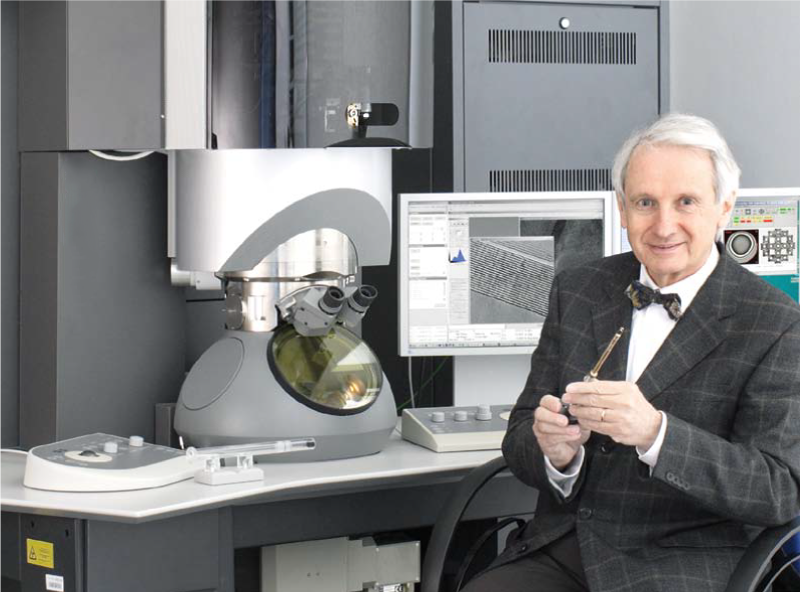
Co-author – Dr. Xing Huang, ETH Zürich

Learn more about our Climate system and Nano-Reactor:

Discover more publications made possible by our Climate system:

Interview with Prof. Rafal Dunin-Borkowski, Director of Ernst Ruska-Centre for Microscopy and Spectroscopy with Electrons in Jülich

Interview with Prof. Rafal Dunin-Borkowski, Director of Ernst Ruska-Centre for Microscopy and Spectroscopy with Electrons in Jülich

Fig. 1. Prof. Rafal Dunin-Borkowski. Photo credit: Forschungszentrum Jülich

We interviewed Rafal Dunin-Borkowski, Director of Ernst Ruska-Centre (ER-C) for Microscopy and Spectroscopy with Electrons in Forschungszentrum Jülich. We talked about his road to ER-C, his research into more energy-efficient electronic devices, the growing importance of software and data analysis and the need for automation to improve the measurement of weak signals. 

“I currently have the greatest personal interest in developing techniques for characterizing the functional properties of working electronic and spintronic devices on the smallest scale and in real time in the presence of stimuli such as applied field, voltage, temperature, light, gases and liquids.”

Where does your passion for Electron Microscopy come from?

My passion for electron microscopy was accidental. It came from being taught by Michael Stobbs as an undergraduate and during my Ph.D. He communicated his enthusiasm for developing and applying characterization techniques as a combination of fundamental physics, materials science and other scientific disciplines. Almost every problem involves exploring a new material or phenomenon at close to the atomic scale that no one has studied before.

Can you tell us about your road to the Ernst Ruska-Centre in Jülich?

It was a long road! First of all, I was in Cambridge University, where I completed my undergraduate degree in physics, my Ph.D. and first postdoctoral appointment with Michael Stobbs. I then went to Arizona State University, where I was sponsored by IBM Almaden and worked with David Smith and Molly McCartney on magnetic recording technology. In Arizona, I also worked with John Cowley, Peter Buseck and Michael Scheinfein. I then went to the Department of Materials in Oxford University for 2 years, where I was responsible for using a new field emission microscope with internal and external users. I then obtained a Royal Society University Research Fellowship, returned to Cambridge University and stayed there for almost 7 years, working primarily on off-axis electron holography and related techniques. After Cambridge, I was employed in the Technical University of Denmark to set up a new department, which was called the Center for Electron Nanoscopy. I stayed there for 5 years.

Fig. 2. Prof. Knut Urban. Photo credit: Forschungszentrum Jülich

In 2011, I took over the Institute for Microstructure Research in Forschungszentrum Jülich in Germany when the previous director, Knut Urban, retired. This institute has a long history in electron microscopy technique development and applications, as well as in the operation of the Ernst Ruska-Centre as a user facility. Together with colleagues in Heidelberg and Darmstadt, Knut Urban contributed to the development of spherical aberration correction for transmission electron microscopy in the 1990s. Forschungszentrum Jülich has been operating the Ernst Ruska-Centre as an international user facility since 2004, together with RWTH Aachen University. 50 % of the access time to the instruments is made available to external users, who work with our experienced scientific and technical staff.

As a director, are you still involved in hands-on research?

In the institute that I direct in Jülich, we currently have about 100 active scientists and students, many of whom are paid from 3rd party funding. This means that we respond to external funding decisions, which determine the scientific directions that we work on. It also means that I spend a lot of time raising funding or managing research projects. I therefore have little time to do hands-on research myself. However, I try to stand behind people when they use the electron microscopes and help them with writing software and data analysis. In addition, if any research paper has my name on it I try to make sure that I comment on it line by line. In this way, I try to take as active a role in scientific research as I can.

Which were defining moments that accelerated your career?

Scientifically, there were certain people I worked with who were very helpful in my development
as a scientist. In particular, working with Michael Stobbs, David Smith, John Cowley and others gave
me key experiences and insight. Now, I try to facilitate an environment for people to do the kind of
work that I would like to be doing myself. I look forward to not being a director and going back to
doing hands-on research in the future, because I regard this as my strength.

Fig. 3. Members of the ER-C team (from the left): Dr. Karsten Tillmann, Dr. Juri Barthel, Marita Schmidt and Dr. Andreas Thust.
Photo credit: Forschungszentrum Jülich

What makes the ER-C a unique institute?

The Ernst Ruska-Centre is unique in many ways. It is managed both from the Jülich Research Center and from RWTH Aachen University. This means that there is frequent interaction between people who work in both places, as well as with external users of the facility. We encourage external users to come for as long as possible, so that they are genuine collaborators with our research staff, who each have their own research topic to work on. We also try to encourage our staff to work on technique and instrumentation development to tackle new problems that are brought to us.
The Ernst Ruska-Centre is now moving from research only in the physical sciences to also include soft materials and life science. This change in the breadth of our research allows us to apply techniques, instrumentation and software that have been developed to tackle problems in the physical sciences to soft and biological materials, and vice versa. We are also establishing closer links with other characterization techniques, especially neutron science and synchrotron X-rays, as well as with data scientists.

Fig. 4. Forschungszentrum Jülich – Staff. Photocredit: Forschungszentrum Jülich

What is the role of the ER-C on a global scale?

On a global scale, at first sight the Ernst Ruska-Centre resembles how user facilities work elsewhere, for example in the US National Laboratories. In practice, the working principle is different, in particular with regard to the fact that all of our staff work on as long-term a collaborative basis as possible with incoming scientists and students, in order to optimize experiments and data analysis together with them, rather than concentrating on serving many users.

Do you collaborate with industry to develop new techniques?

In the ER-C, we try to go beyond the techniques and capabilities that are available elsewhere, for example by undertaking ambitious development projects with manufacturers, where we commit our staff time in return for access to technology that is not yet available commercially. Software and instrumentation that is developed in the ER-C is then often licensed back to the manufacturers for the benefit of their future customers and the community as a whole.

In which research topics are you personally interested?

We currently have more than 10 working groups in the ER-C, many of which focus strongly on technique development, as well as on specific materials problems. I have an interest in almost every activity in the institute.

Fig. 5. Artist impression of Spintronics.

However, I currently have the greatest personal interest in developing techniques for characterizing the functional properties of working electronic and spintronic devices on the smallest scale and in real time in the presence of stimuli such as applied field, voltage, temperature, light, gases and liquids. Many of these capabilities have only recently become available. The experiments are carried out at the highest spatial resolution using phase contrast and spectroscopic techniques in both TEM and STEM imaging modes. They also require the development of new approaches for handling the increased amount and rate of data coming from the microscopes.

To what extent do societal challenges determine your choice in your research topics?

Societal priorities have a decisive factor on which scientific topics are funded. In turn, they drive our research. In the Helmholtz Association, we work on the basis of program oriented funding. Every 5 years, our scientific priorities are redefined, in part by societal needs. At the same time, by its very nature much of our research is exploratory and operates over longer timescales, especially with regard to technique and instrumentation development.

Will in situ techniques play a role in the research of ER-C and why are these in situ techniques becoming relevant?

A variety of different problems come under the heading of in situ electron microscopy. Some of our experiments involve “in situ” chemical reactions in gas or liquid environments, while others involve passing electrical currents through or applying magnetic fields to nanoscale materials, or studying the effect of temperature, light or mechanical stress.

One of the scientific priorities of the Hemlholtz Association, which funds much our research, is to understand and develop more energy-efficient devices for future computing applications. In our institute, we use electron microscopy to map the local crystallography, microstructure and functional properties of novel nanoscale devices in real time. We would like to make these measurements on ever faster timescales and are currently developing new hardware and software that we hope will give us access to the sub-nanosecond regime.

What do you expect from DENSsolutions in the future?

We have a partnership agreement and many specimen holders from DENSsolutions, which we are very pleased with. We would like to have an even closer partnership in the future and have many ideas for more ambitious technical developments, as well as for the automation of complicated workflows. In particular, the current practice of performing experiments manually limits our ability to measure very weak signals, which would require repeating the same sequence of steps many thousands of times. For this reason, we need the kind of automation of experiments that is now available in the life sciences. We understand that there is a greater variety of samples and experiments in the physical sciences and that such workflows would then have to be more flexible.

Is your goal with automation to get a higher throughput for your experiments?

This is not the priority. I would primarily like to use automation to improve the measurement of weak signals and to obtaining better statistics in certain measurements, rather than simply to achieve high throughput. We therefore also need more stable specimen stages and a cleaner environment in the microscope column so that the sample does not change over time. There is one other aspect of automation that does not exist at the moment, which is the ability to store samples, for example in inert environments in individual cartridges, until they are no longer needed, perhaps over many years, so that the same region of the same sample can be reassessed quickly, easily and reproducibly as many times as required.

Want to read more?

Did you like this interview? Subscribe to our newsletter to receive more in situ related news like this.

Or follow us on social media:

Real‐Time Imaging of Nanoscale Redox Reactions over Bimetallic Nanoparticles

Real‐Time Imaging of Nanoscale Redox Reactions over Bimetallic Nanoparticles

Shu Fen Tan, See Wee Chee, Zhaslan Baraissov, Hongmei Jin, Teck Leong Tan, Utkur Mirsaidov
Published in Advanced Functional Materials on 16 July 2019

Publication

Video 1: In situ TEM movie of a Pt–Ni rhombic dodecahedron NP undergoing oxidation reaction in 20% O2 / 80% N2 gaseous environment at 400 °C inside a gas cell.
Video 2: In situ TEM movie of a Pt–Ni rhombic dodecahedron NP undergoing reduction reaction in 5% H2 / 95% N2 gaseous environment at 400 °C inside a gas cell.

This recent article from National University of Singapore about the catalyst activity of bimetallic nanoparticles clearly demonstrates the versatility of the Climate G+ gas & heating system that was used for the in situ TEM experiments.

Figure S4. In situ TEM images showing the evolution of Pt–Ni NPs (A) before and (B) after the oxidation (20% O2 / 80% N2) reaction followed by (C) the reduction (5% H2 / 95% N2) reaction at 400 °C inside a gas cell. Here, t = 0 s marks the start of 20% O2 / 80% N2 flow. At t = 133.1 s, the gas mixture was switched to 5% H2 / 95% N2.

Fast switching between oxidizing and reducing gas conditions allows direct observation of the morphological changes in the catalyst material. The video’s above also demonstrate the UHR image stability at in situ conditions.

Combining the images and video’s from this experiment with SAED, EDS and Mass Spectroscopy results gives valuable information about the areas where the catalyst is most active.

Nanoscience Instruments and DENSsolutions announce new partnership

Nanoscience Instruments and DENSsolutions announce new partnership

DENSsolutions is proud to partner with Nanoscience Instruments to serve the US and Canadian markets. Nanoscience Instruments combines expertise in microscopy and surface science instrumentation with real-world solutions. This partnership will provide a synergistic fit to the core competencies of Nanoscience Instruments and the markets they serve.

Sebastian Kossek, co-owner of Nanoscience Instruments

“The DENSsolutions In Situ TEM solutions allow researchers to fully investigate real-world phenomena and achieve breakthrough science. The expansion to our range of products will further expand our reach into key strategic markets,” says Sebastian Kossek, co-owner of Nanoscience Instruments.

“As a company that specializes in surface science characterization and measurement instrumentation solutions, we are excited to have additional tools that are synergistic to our current and future customer base.”

“As scientists and researchers are looking to solve macro modern-day global challenges which society is facing, they need to understand materials down to the atomic level. The innovative and powerful In Situ TEM solutions DENSsolutions provides helps these researchers change the world, one atom at a time,” says Ben Bormans, CEO DENSsolutions.

“By incorporating all stages of the development model, In Situ TEM unlocks unprecedented research capabilities and moves nanoscience to the next level.”

Ben Bormans, CEO of DENSsolutions

Ben Bormans, CEO of DENSsolutions

“As scientists and researchers are looking to solve macro modern-day global challenges which society is facing, they need to understand materials down to the atomic level. The innovative and powerful In Situ TEM solutions DENSsolutions provides helps these researchers change the world, one atom at a time,” says Ben Bormans, CEO DENSsolutions.

“By incorporating all stages of the development model, In Situ TEM unlocks unprecedented research capabilities and moves nanoscience to the next level.”

Mark Flowers, co-owner of Nanoscience Instruments.

“The partnership with DENSsolutions opens new avenues for the current customer base and find additional overlaps with the other technologies we currently provide,” says Mark Flowers, co-owner of Nanoscience Instruments.

“Observing processes ‘on-site’ as they are occurring and under changing external stimuli is the paramount goal of In Situ, time resolved techniques.”

For more information about Nanoscience Instruments and the products and services they provide, visit us online at www.nanoscience.com

Interview with Prof. Sara Bals, head of EMAT Antwerp

Interview with Prof. Sara Bals, head of EMAT Antwerp

We interviewed Sara Bals, head of the Electron Microscopy group at the University of Antwerp (EMAT). We talked about her passion for electron microscopy, her team and the importance of tomography when creating 3D images that reveal the real structures of nanoparticles and clusters. This technique can lead to the development of novel materials and improvements in application fields such as catalysts.

“The idea that we can see what nobody else can see, that today might be the day when we discover something novel, that is really what keeps me motivated and what keeps me going.”

Where does your passion for Electron Microscopy come from?

When I had to choose a topic for my master’s thesis, I decided to start working here at EMAT with Professor van Tendeloo and he introduced me to the basic principles of transmission electron microscopy. During my master’s thesis I used this technique to investigate superconducting thin films and tapes, and I continued this research during my PhD. After my PhD, I went to the National Center for Electron Microscopy in Berkeley and there, I discovered the possibility of investigating nanomaterials in three dimensions using electron tomography.

Fig. 1. DENSsolutions Wildfire Tomography holder

Tomography is based on the acquisition of a tilt series of two-dimensional projection images. You use those images as an input for a three-dimensional reconstruction. I became really passionate about this technique because without this every image that you acquire using a transmission electron microscope is only a two-dimensional projection of a three-dimensional object and that can be very misleading.

What I like about electron tomography is that it is a very visual technique. Once the reconstruction is obtained it is rather straightforward to interpret the results. And the idea that we can see what nobody else can see, that today might be the day when we discover something novel, that is really what keeps me motivated and what keeps me going.

Can you give an example of one of these discoveries?

In 2018 We received samples from the group led by Professor Sara Skrabalak at Indiana University. She sent us samples where, from two-dimensional projections, we could see that there was some sort of structure. However, when we performed the electron tomography you could see that these particles were extremely symmetrically shaped octapods1. So the idea that you can visualize that, and that you can give feedback to the growers of these materials showing them what they have made, that is really very nice.

Fig. 2. Nano dumbbells. Nano Lett. 2012 Aug 8;12(8):4380-4. doi: 10.1021/nl3021957. 

Also, something that we have been investigating quite a lot recently are assemblies of nanoparticles. These are groups of nanoparticles of which from a two-dimensional projection image you can not say how many particles you have in the assembly or how they are organized. So we did tomography on one of these assemblies from the group of Professor Luiz Liz-Marzán who is the director of the CIC biomaGUNE Institute in San Sebastian. And we could see that this assembly was almost a perfect three-dimensional puzzle of what is called gold nano dumbbells (Fig. 2.).

Developing electron tomography at high temperatures2was one of the aims within the EUSMI project where EMAT, Denssolutions and CIC BiomaGUNE are partners. It is great to see these groups joining forces.

What is the promise of this research into gold nanoparticles?

They can be used for several applications; they can be used as sensors or they can be used for medical applications or it can be used for catalysis. So there is a broad variety of applications but very often the properties of these nanomaterials; the catalytic properties, the optical properties, depend on the three-dimensional shape. That’s why it’s important to investigate the shape and the three-dimensional structure using electron tomography and we want to do that with atomic resolution because you can consider nanoparticles to be agglomerates of individual atoms.

So if we are able to determine the positions of the individual atoms, together with their chemical nature and the bonding between them, then we can provide the necessary input to predict their properties through simulations. In this manner we may even guide the synthesis of novel nanomaterials.

Fig. 3. The research team of Sara Bals at EMAT

What makes your research group, EMAT, unique in the world of Electron Microscopy?

EMAT is the electron microscopy group at the University of Antwerp. We are quite a large group. We have about 60 researchers. At the moment we have six transmission electron microscopes of which two are aberration-corrected. But I think it’s not the instruments that make us unique. It’s really the team and I often say to new people or people I’m talking with: ‘team’ has the same letters as EMAT because we work together as a team.

At EMAT we have different principal investigators. They all have their own expertise but we work together and the expertise is very complementary. That is what I enjoy most about my work; that we can share knowledge and combine knowledge and that we can create bigger results than what we would be able to do on our own. So the research that we are doing is a good mixture of fundamental, applied and industrial research. But in any case, our main goal is to solve relevant problems in materials science.

Can you give us some examples of these relevant problems that you want to solve?

For example to really understand the connection between the properties and the structure of materials. So we’re not just trying to improve the record of resolution in a well-known material. Let’s say silicon. We want to investigate real-life materials that are sometimes difficult to image because they’re electron beam sensitive or they have many different types of elements. Those are the kinds of materials that we want to investigate. I also think about metallic nanoparticles with the applications that I’ve already mentioned, or maybe some of the organic perovskites.

We’re developing novel techniques in collaboration with people making these samples and we try to understand very well what the questions are that they would like to answer. This work is more challenging but more meaningful and it really pushes the boundaries of technology. Projects that have this combination are my favourites.

How do the in situ stimuli provided by the DENSsolutions systems, like gas and heating, contribute to your research?

Recently I realized that all of the experiments we’ve done so far are done under the conventional conditions of an electron microscope meaning room temperature and ultra-high vacuum. And those conditions are no longer sufficient if you want to understand the behaviour of these nanoparticles and their applications. So that’s why we started thinking; can we visualize the three-dimensional changes that these nanomaterials undergo when they’re exposed to high temperatures or high pressures? So that is what our main goal is within my ERC consolidator grant REALNANO where we are trying to combine the principles of electron tomography with in situ electron microscopy.

So far we got some preliminary results using the Climate gas system, investigating platinum nanoparticles. These are of importance for catalytic applications. But we know that the surface structure might change in a gaseous environment. So this is what we wanted to investigate.

Again we want to perform these measurements in three dimensions and not just based on a two-dimensional projection of a three-dimensional object which can be very misleading. So by combining the expertise of several of the principal investigators here within the EMAT group and with the help of DENSsolutions, we were able to do some first experiments where we acquired high-resolution images of these platinum nanoparticles.

From these images, we’re going to quantify how many atoms we have in a given atomic column and these counting results serve as an input for molecular dynamic simulations that enable us to obtain a three-dimensional model.

Fig. 4. DENSsolutions Climate Gas supply system

Fig. 5. DENSsolutions Climate Gas analyzer

Now using this methodology we investigated a given nanoparticle. We looked at that nanoparticle in a vacuum, in hydrogen and in oxygen. Using the DENSsolutions Climate gas supply system we kept on switching between hydrogen and oxygen. What we saw is that in hydrogen clear facets are present whereas in oxygen a more round structure is observed. And so these surface dynamics are very important if later want to understand what happens to these particles during catalytic reactions. So we have been developing the methodology and we really look forward to investigating these systems under real catalytic conditions.

Thanks to a recent ERC consolidator grant we were able to acquire our own Climate system. Using this system, also in combination with the included Gas Analyzer, we are planning on expanding this type of research.

Can you elaborate on the future investigation under real catalytic conditions?

We want to start investigating different sorts of catalytic nanoparticles, not only model-like systems. For instance; supported nanoparticles or hetero nanostructures rather than one type of element. There are plenty of experiments and different catalytic reactions we can think of plenty of so we’re also collaborating with a lot of groups in Europe to figure out what would be the most important tests that we could perform in this with the Climate system. And of course, we’re also trying to continuously improve our methodologies to visualize the three-dimensional structure.

This is also why we’re really happy that there is currently a heating tomography holder available which tilts across a range of plus-minus 70 or 80 degrees. This is the holder that we have used to investigate the thermal stability of gold stars and octapods in the past.

EMAT also recently acquired a Stream, liquid and biasing, system from DENSsolutions. What are your plans with this?

My colleague Professor Joke Haderman is investigating battery materials under realistic, in situ, conditions.

We also would like to investigate assemblies of nanoparticles in a liquid state. Because these assemblies are formed in a liquid, creating a three-dimensional structure. Then if we would put this structure on a conventional TEM grid there is a possibility that the shape will deform. So far we have been investigating all of them in three dimensions but in a dried state and what we could try and figure out now is how different this dried state is from the native state in a liquid. So we have been doing some very preliminary testing on that and I have a postdoc who is going to apply for a project in order to obtain funding to really push this research.

Next to your current projects, is there any other research at the moment that excites you?

What I am really interested in is the fact that all of these nanoparticles, or at least the nanoparticles that I’ve been investigating most: colloidal nanoparticles, are covered by surface ligands and most of the time when using an electron microscope we completely ignore these. Surface ligands maintain the shape of the nanoparticle but they also form the interface with the environment so they are extremely important.

We do not visualise them because they contain very light elements such as carbon. This makes imaging more complicated because sometimes they form a carbon shell. But I started to understand that it is very important to visualize them because they will interact with the environment first. So we have been looking into how to visualize these surface ligands using more advanced electron microscopy techniques. Also by exploiting the single electron detectors that nowadays became available and also by looking into the support that we are using in the transmission electron microscope. So using graphene type supports rather than the conventional carbon supports. And so when I heard that also DENSsolutions is thinking in that direction I was also very excited about the idea of replacing the silicon nitride by the graphene and I think that again there will be a lot of new possibilities through such chips.

Fig. 6. Graphene – artist impression

I think the graphene support would make a lot of people happy and increasing the tilt capabilities of a Climate system would also open up a lot of possibilities for new experiments, especially in the field of catalysis. Think about supports loaded with catalytic nanoparticles where you really want to understand the three dimensional structure and understand for example degradation mechanisms in three dimensions.

How do you experience your collaboration with DENSsolutions?

Well, something that I really appreciate is that for example at the moment one of my students is performing an internship at DENSsolutions. This is in the framework of a Marie Curie training network called Mummering. So I think it’s really great that DENSsolutions is offering him the possibility to do a secondment there because I believe that once he understands how the chips are made and what the ideas behind the approach are that this will enable him to perform electron tomography experiments at high temperature in a much more efficient thought through manner. So this is really something that I appreciate that DENSsolutions also wants to put the effort into training students like that.

I also appreciate that when we are doing an experiment where we think: OK this is it, now everything is coming together, we have the right samples and we know what we want to aim for, that during the experiments that we did, which later ended up in the Nanoletters paper3, that people from DENSsolutions came over to help and we could do the experiments together. This really accelerated the experiment which is important because of microscopy time is valuable. So it was really nice that it was a group effort into getting those results.

Did you like this interview? Subscribe to our newsletter to receive more in situ related news like this.

Scientific references

1 “Thermal Stability of Gold/Palladium Octopods Studied in Situ in 3D: Understanding Design Rules for Thermally Stable Metal Nanoparticles” Albrecht W, Bladt E, Vanrompay H, Smith J.D,Skrabalak S.E, Bals, S, ACS Nano 13, (2019) 6522-6530

2 “3D characterization of heat-induced morphological changes of Au nanostars by fast in situ electron tomography”. Vanrompay H, Bladt E, Albrecht W, Béché A, Zakhozheva M, Sánchez-Iglesias A, Liz-Marzán LM, Bals S, Nanoscale 10, 22792 (2018)

3 “Three-Dimensional Quantification of the Facet Evolution of Pt Nanoparticles in a Variable Gaseous Environment”. Altantzis T, Lobato I, De Backer A, Béché A, Zhang Y, Basak S, Porcu M, Xu Q, Sánchez-Iglesias A, Liz-Marzán LM, Van Tendeloo G, Van Aert S, Bals S, Nano Letters 19, 477 (2019).