Izasa Scientific and DENSsolutions announce new partnership

Izasa Scientific and DENSsolutions announce new partnership

DENSsolutions is proud to partner with Izasa Scientific to serve the Spanish and Portugese market. We have been active in Spain for a number of years and in those years we started to build a good relationship with the expert and passionate team of Izasa Scientific. Now we have recently officially signed our partnership, which we are sure will benefit the TEM community in Spain and Portugal. Offering them cutting-edge In Situ technology with the best possible local support.

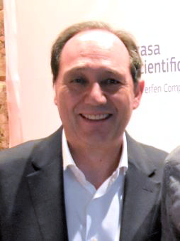
“During conversations and cooperation with DENSsolutions on specific projects in Spain, we became convinced about the perfect alignment between our companies. The technology offered by DENSsolutions fits perfectly with our product portfolio linked to Electron Microscopy. Now, we can offer our clients a complete on-site solution by introducing the DENSsolutions systems that complement the most advanced Electron Microscopes and direct acquisition cameras on the market.
We are convinced that the association between our companies will end in a clear benefit for the scientific community by facilitating access to the most complete solution for “in-situ” Electron Microscopy on the market. We believe that DENSsolutions is without a doubt the best partner in this field.
This good alignment, even before formalizing this agreement, which we are pleased to announce, has led to joint efforts, such as the recent webinar, which attracted considerable interest from the scientific community, and which we invite you to watch on the Izasa Scientific website.”
Carlos Arribas, General Manager at Izasa Scientific
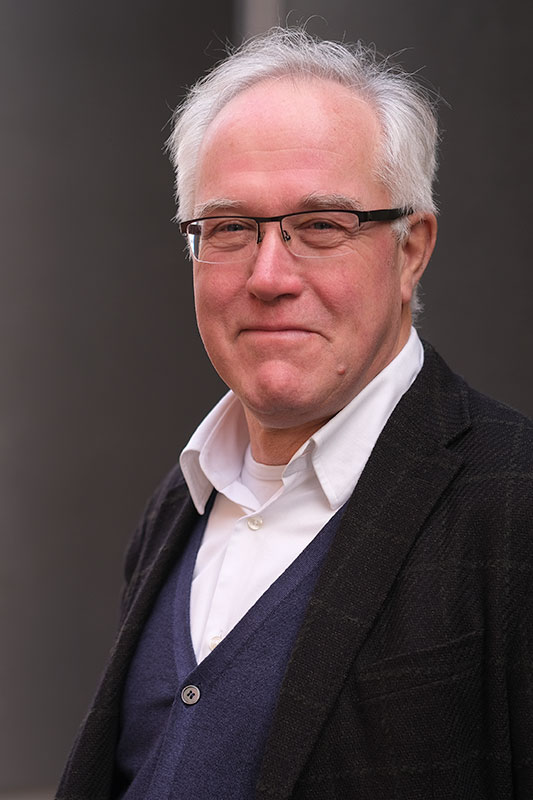
“In the last 5 years, DENSsolutions has had the opportunity to deliver in situ heating and biasing solutions to some of the leading TEM laboratories in Spain. We also saw more and more requests for quotations for our Climate and Stream systems so we decided to start looking for a distributor in Spain and Portugal. We already had some contact with Izasa Scientific in a few projects and we got very impressed with their installed base and more importantly with the commitment of the employees of Isaza towards their customers. The core values of DENSsolutions are: “we care”, “we innovate” and “we deliver” and this is exactly what we found with the people in Izasa.
So I fully agree with Carlos Arribas, the General Manager of Isaza Scientific, when he states that there is good alignment between the two companies. It was clearly shown indeed during our joint recent webinar.
DENSsolutions will keep pushing the technology envelope for total in situ solutions based on our state of the art Nano-Chip, Nano-Reactor and Nano-Cell MEMS based sample carriers.
In order to support our customers in every part of the world, we believe that a true partnership with our distributors is extremely important and after careful consideration we decided that this true partnership for Spain and Portugal can be realized with Isaza Scientific in the most beneficial way for our customers.
Isaza Scientific brings a lot of value for DENSsolutions as they have a very extensive installed base in Spain and Portugal. But more importantly the people in Isaza Scientific really have the knowledge and drive to understand our customers’ needs.
Last but not least: with the completion of the distribution agreement with Isaza Scientific we now “cover” the whole of Europe.”
Ben Bormans, CEO -DENSsolutions

Do you want to receive great articles like this in your mailbox? Subscribe to our newsletter.

In Situ helps to understand the recovery of deactivated palladium catalyst

In Situ helps to understand the recovery of deactivated palladium catalyst

In Situ images reveal that nanoparticles can be transformed into more active atomic species at high temperatures

Original article by Huang Zhou, Yafei Zhao, Jie Xu, Haoran Sun, Zhijun Li, Wei Liu, Tongwei Yuan, Wei Liu, Xiaoqian Wang, Weng-Chon Cheong, Zhiyuan Wang, Xin Wang, Chao Zhao, Yancai Yao, Wenyu Wang, Fangyao Zhou, Min Chen, Benjin Jin, Rongbo Sun, Jing Liu, Xun Hong, Tao Yao, Shiqiang Wei, Jun Luo & Yuen Wu. Published in Nature Communications volume 11.

Supported metal catalysts have important applications in many industrial processes like the production of chemicals, pharmaceuticals and clean fuels, and the purification of vehicle emissions. At elevated temperatures the small catalyst particles tend to form bigger particles due to a process called sintering, which decreases their active surface areas and diminishes the catalytic activity. Replacing the deactivated metal nanoparticles is a costly process. Therefore researchers are looking for ways to improve the sustainability of these catalysts. In this study, scientists from multiple Chinese institutes, including DENSsolutions customer Tianjin University of Technology, further researched a method to recover or regenerate the activity of sintered and deactivated catalysts.

Figure 1a. Clusters of Pd nanoparticles, as seen in the upper left picture, are thermally diffused in N-doped carbon layers at 900 °C under Ar atmosphere – images taken from a video. Scale bar, 5 nm.

Figure 1b. Detailed view of Pd single atoms in the N-doped carbon layers after the in situ observation in Figure 1a.

Findings

In this in situ experiment the researchers discovered that supported palladium/gold/platinum nanoparticles distributed at the interface of oxide supports and nitrogen-doped carbon shells would undergo an unexpected nitrogen-doped carbon atomization process against the sintering at high temperatures, during which the nanoparticles can be transformed into more active atomic species.

In Situ TEM study

In order to study the thermal diffusion of the Pd nanoparticles within N-doped carbon layers, a sample environment with an inert gas like Argon needs to be created. The big advantage of the Climate system is that it can create this sample environment inside a normal TEM without the need of an ETEM. Furthermore, the high stability of the Climate Nano-Reactor allowed the researchers to record the N-doped carbon atomization process with sufficient detail in order to get valuable insights.

Figure 2. Representative in-situ TEM images of Pd NPs/TiO2@PDA-Pd NPs/TiO2@C. (a-h) Different temperatures, (i-l) different times at 900 °C. Scale bar, 20 nm.

“Thanks to the wonderful gas cell system from DENSsolutions, we can directly observe and record the sintering process of metal catalysts from 100 to 900 °C under 1 bar Ar by transmission electron microscopy (TEM).
We also succeeded in tracking the N-doped carbon atomization process and the evolution of metal nanoparticles into metal single atoms. During these processes, the response of this gas cell system was very fast and perfectly stable.”

Prof. Jun Luo
Professor at the Center for Electron Microscopy, Tianjin University of Technology.

Learn more about our Climate system:


Discover more publications made possible by our Climate system:


Read the original article:

Do you want to receive great articles like this in your mailbox? Subscribe to our newsletter.

Improved FIB lamella preparation

Improved FIB lamella preparation

A conversation with our Product Manager Dr. Yevheniy (Gin) Pivak on the new FIB stub 3.0

DENSsolutions introduces the 3rd generation of the FIB stub which enables researchers to prepare a lamella and place it directly on the Nano-Chip, all inside the FIB. In this version, many improvements were made to make the FIB sample preparation easier, safer and quicker. The development of this new stub was headed by our Product Manager Dr. Yevheniy (Gin) Pivak in close collaboration with key partners like AEM, TU Darmstadt and EMAT, Antwerp.

Why was this new FIB stub designed?

Any TEM experiment starts with a good sample either it’s a nanoparticle, a FIB lamella, 2D material or a nanowire. The FIB sample is the most complicated among others, especially when it comes to preparing lamellas onto MEMS-based Nano-Chips.
Several years back, when the users’ knowledge on FIB lamella preparation onto MEMS-based Nano-Chips was still very limited and the field itself was premature, DENSsolutions developed the first version of a FIB stub which had two inclined sides of 45 degrees on which the sample and Nano-Chip could be positioned. This stub simplified the sample preparation process by allowing the user to prepare and transfer a lamella onto the Nano-Chip in one go without breaking the vacuum of the FIB chamber, thus saving operation time. Since then version 2.0 was released, which kept the main design features like the two inclined sides but improved the sample and Nano-Chip positioning and clamping.
In the meantime, hundreds of lamellas were successfully prepared and placed onto Nano-Chips but many users still encounter challenges during the process. First of all, the issues come from an uncommon geometry that the users need to work with; the lamella preparation and the lift out need to be done at 45 and 55 stage tilt angles. On top of that, the users suffer from poor imaging, especially at low accelerating voltage and a charging effect. The positioning of the Nano-Chip and the clamping mechanism, that is also there for grounding purposes is not optimal, making the operation not very user-friendly. Because the height of the sample and the Nano-Chip on the FIB stub can differ quite a bit, there also is a safety concern.
In recent years more and more people are interested in in-situ TEM Biasing and Biasing & Heating experiments. The majority of those samples are FIB lamellas and the requirements towards the samples for electrical measurements are much stricter compared to heating experiments with various pitfalls along the way. A new approach aiming to avoid short circuiting and reiteration of biasing and biasing & heating lamellas is required.

What are the benefits of this new FIB stub?

The new FIB stub solves a number of limitations of the previous versions.

At first, the new 3.0 stub incorporates an additional flat side for placing the samples that ensures a conventional geometry and the very same and the well-known process used by any FIB operator when making and lifting out the lamella.

The revised geometry improves the quality of imaging even during the low kV milling and polishing steps. Additionally, the charging effect is reduced due to a more effective grounding of the Nano-Chips’ contact pads.

A dedicated pocket and a smart clamping mechanism is introduced which drastically simplifies and speeds up the Nano-Chip loading and unloading, making it very user-friendly. It reduces the risk of breaking the membrane when handling the Nano-Chips and there is also no need to use sticky tapes to fix or to ground the Nano-Chip, which in turn makes the process a lot cleaner.

The design of the FIB stub brings the position of the sample and the Nano-Chip to a similar eucentric height, minimizing the possibility of crashing into the pole piece, the Gas Injection System or the manipulator during the operation.

What is the compatibility of the new FIB stub?

The new FIB stub is compatible with Thermo Fisher/FEI and JEOL dual beams. It’s suitable for various models like Strata DB235 (Thermo Fisher/FEI), Helios NanoLab 600 / 650 / G4 CX (Thermo Fisher/FEI), JIB 4600F (JEOL) and many more.
It’s also suitable for any Thermo Fisher (FEI) and JEOL double tilt Heating and/or Biasing Nano-Chips.

Who are the people that will benefit from it?

Any existing customers who own a double tilt Wildfire TF(FEI)/JEOL (Wildfire D6, Wildfire H+ DT), a Lightning HB TF(FEI)/JEOL (Lightning D6+) or a Lightning HB+ TF(FEI)/JEOL (Lighting D7+, Lightning D9+) system and works with FIB lamellas will definitely benefit from the new FIB stub.
New customers of Wildfire and Lightning systems planning to work with in situ heating samples or electronic devices like non-volatile memory based on resistive switching or phase change materials, solid state batteries, solar cells, etc will enjoy the sample preparation using the 3.0 stub

What kind of challenges were tackled during development?

As in many developments, the main goal is to create a really good product that can be used by most of the users. However, because there are many dual beams from different manufacturers out there with their own stage and column design, various manipulators, workflows, details, etc. that also can vary from site to site it is quite challenging to make one generic product that is suitable for everybody. It’s not possible to fulfil everyone’s needs, but we spent a lot of time trying to get the new FIB stub as versatile as possible.
In any case, product development is a dynamic process. As long as researchers find bottlenecks in their pursuit to get the right research results, we will focus our efforts to provide them with the right solution.

Did we cooperate with customers on this development?

Any product is meant to solve customers issues and limitations or create new opportunities. We make products for our customers and not for ourselves and there is no way to make a good product without customers involvement.
Following our strategy, we involved a number of our close collaborators during the development and testing of the new FIB stub project, namely EMAT (University of Antwerp) and AEM (University of Darmstadt). Additionally, more customers from Germany, UK, Singapore, Spain, Sweden, etc. were involved in the initial discussion phase to identify the current issues and limitations with the FIB lamella preparation.

Which future developments lie ahead?

In the near future, the intention is to verify the compatibility of the FIB stub 3.0 in Zeiss, Tescan and Hitachi dual beams.
If you are a proud owner of one of above-mentioned FIBs and you would be interested to test the new stub, please contact us.
On a longer term, we are working to further improve the electrical quality of lamellas and devices prepared on biasing and heating and biasing Nano-Chips. This next development is planned to be present at the EMC 2020 conference in Copenhagen. So, stay tuned!

5 reasons to get the new FIB stub:

Receive a quotation:

Do you want to receive great articles like this in your mailbox? Subscribe to our newsletter.

First Visualisation of Crystal Growth from Organic Molecules

First Visualisation of Crystal Growth from Organic Molecules

Scientists characterise how flufenamic acid, a COX inhibitor, which is used in multiple industries, forms crystals from a liquid solution

Original article by Jennifer Cookman, Victoria Hamilton, Louise S. Price, Simon R. Hall and Ursel Bangert. Published in Nanoscale, issue 7, 2020.

The earliest stages of crystal growth are key to determining the final structure of a crystal, and scientists have visualised this process for the first time. Organic molecules were formed and grown in a liquid environment and characterised using the DENSsolutions Ocean In Situ system for Liquid Phase Electron Microscopy (LPEM).

Experimental Firsts

“Using LCEM (liquid cell electron microscopy) it has been possible for the first time to capture early-stage nucleation events of organic molecular crystals used as APIs (active pharmaceutical ingredients),” said Dr Jennifer Cookman from the Bernal Institute at the University of Limerick and lead researcher for this study.

“The importance of this method is that we can begin to understand how one crystal structure forms over another and, even more importantly, how these early-stage nucleation events manifest. We can also compare/contrast with classical nucleation theory and other crystal growth theories.”

The molecule studied, flufenamic acid, is a COX-inhibitor that acts as an anti-inflammatory. With increased study of this molecule, which is used in the pharmaceutical industry, researchers can potentially fine-tune its action as a medicinal drug and reduce its side-effects.

What’s more, the molecular crystalline state is widely used across many industries; including electronics and agrochemicals. This research represents the first steps to analysing molecular crystal growth of not just flufenamic acid, but other molecules with implications for improvements in other industries.

A 55-second video of flufenamic acid crystal growth at a scale of 0.5 micrometers. This video is remarkable as it shows the entire process of crystal nucleation, from a blank screen through nucleation to crystals. A good region to examine is the central point just above the 0.5 μm scale. A crystal forms here and grows without getting cluttered by other crystals.

Techniques and Methods

The Ocean LPEM system’s unique ability to perform transmission electron microscopy in a liquid ethanol environment was essential for this research. Dr Cookman adds that by “using the DENSsolutions Ocean holder we were able to introduce an undersaturated liquid solution of the API to be visualised in the TEM protected from the high vacuum environment of the TEM.” Then crystal growth was induced by illuminating the sample with an electron beam which provided the energy needed to prompt the nucleation process.

In situ microscopy far exceeds previous ex situ observations as the team could produce live footage of each stage of crystal growth. Additionally, the in situ technique enabled the visualisation of the nucleation of flufenamic acid molecules in a working environment, a.k.a. ethanol. Performing this analysis in a liquid environment as opposed to a vacuum helped to meaningfully ascertain where and how different physical arrangements of crystal structures occur.

“This work brings focus to the use of electron microscopy and in particular in situ TEM equipment for characterisation,” added Dr Cookman. “That can be of utmost importance to the pharmaceutical industry and also in interim characterisation in pharmaceutical crystal research.”

Micrographs showing crystals forming from flufenamic acid and growing into hexagonal crystals. A 0.2-micrometer scale is used for reference.

Wider Importance

It is at this earliest stage of growth that molecules can exhibit polymorphism; crystal structures that are composed of the same molecules but have different physical arrangements. Understanding how different crystal structures form from the same molecule type is desirable for research that needs nanoscale precision. For example, crystals are commonly used in medicines as a way to deliver active chemicals.

The antiretroviral drug, Ritonavir, which is used to treat HIV/AIDS, was pulled from circulation after it was found to contain a polymorphed version of the active drug. The polymorphed form was less biologically active and did not work as intended. Understanding these early steps in crystal growth is key to fine-tuning processes such as drug delivery.

Future Research

Advances in film technology and TEM allowed for direct observation of the nucleation process and this research represents the potential progress in the field of crystallisation. The results indicate that, with more research, scientists can discern the initial phases of crystal growth. This new technique opens up the field of TEM to visualising other crystallisation pathways, interrogate nucleation mechanisms, and explore new innovations.

The research was part of an EU Horizon 2020 FET-Open project named MagnaPharm which focuses on the magnetic control of polymorphism in pharmaceutical compounds. The team, which includes Dr Ursel Bangert and Dr Simon Hall, intend to continue to characterise flufenamic acid and different growth outcomes under different concentrations.

Learn more about our latest LPEM system ‘Stream’:

Discover more publications made possible by our Ocean system:

Read the original article:

Do you want to receive great articles like this in your mailbox? Subscribe to our newsletter.

Installing the first Climate system in Australia at the University of Sydney

Installing the first Climate system in Australia at the University of Sydney

Standing next to the recently installed Climate G+ system: from left, Keita Nomoto, Lizhuo Wang and Dr. Hongwei Liu from the Australian Centre for Microscopy & Microanalysis

Recently, we celebrated the installation of the first Climate G+ system ever in Australia. For this event we interviewed Dr. Bhatia at the Australian Centre for Microscopy and Microanalysis that oversees the Sydney Microscopy and Microanalysis core research facility at the University of Sydney. Earlier, he was key in the decision to purchase the world’s first Lightning STEM stage which established his relationship with DENSsolutions and ultimately lead to the installation of the Climate G+ In Situ TEM platform.

In this interview, we discussed the research needs of his facility and how they will benefit from the solutions designed and manufactured by DENSsolutions.

Can you tell us a bit about Sydney Microscopy and Microanalysis?

Sydney Microscopy and Microanalysis is the central microscopy facility at the University of Sydney. The University of Sydney is Australia’s first university and regularly ranks in the World’s top 50 universities.

We are a multi-user facility that services both the entire university as well as people from across Australia through the Microscopy Australia access scheme. Microscopy Australia provides access to member universities throughout Australia.

This means that we need to provide highly reliable, flexible solutions as we offer our instruments to users with a broad range of applications and skill levels.

Can you give us some examples of applications that your users are involved in?

Our researchers interests are many and varied. Some of the areas that are relevant to the new Climate system include:

  • Hydrogen generation
  • Methane breakdown as a consequence of global warming
  • Environmental corrosion of metals

It is hard to know what projects it will be used for as many users haven’t even presented themselves yet. But that is the point of buying versatile equipment.

In Australia, it is normal for funding for large equipment purchases to come from research grants. Can you tell us who won the grant to acquire the Climate?

A team from the Chemical Engineering Department working on catalysts were responsible for the bulk of the funding. Their contributions from their grant were topped up by the Australian Centre for Microscopy and Microanalysis (ACMM). ACMM supports the operation of the Sydney Microscopy and Microanalysis facility.

What features of the Climate attracted you to this particular system and how do you see the DENSsolutions in situ system benefitting your research?

As a core facility working with researchers from a range of different fields we needed a system with flexibility to cater to their different interests.
Other features of the Climate that that we found attractive were the ability to interchange parts and the ease with which individual components could be replaced. The ability to perform dynamic mixing of gases provided an added degree of versatility.
All these factors contributed to what we considered to be a future-proof design that best suited our facility and the range of potential experiments of our users.
The ability to investigate dynamic processes and to be able to observe these processes in real-time was also important to us. By being able to observe the entire process takes any guesswork out of the equation and means that we don’t miss any critical steps where changes might occur.

How popular has the system been to date?

The system was only installed in November. So far, we have only had three operators and a technician trained, bearing in mind the Christmas, New Year break. We do however intend to train more operators in the near future and can see the Climate being an important research tool.

In your experience so far, how have you found the Climate system?

The installation process was quite straightforward.
The fact that the software uses the same platform as the Lightning system that we already have abbreviated the familiarisation process. The software itself is very easy to use and the system as a whole is very intuitive.
We have only performed some basic measurements so far, but are looking forward to getting into some detailed experiments in the near future.

Learn more about our Climate system and Nano-Reactor:

Discover publications made possible by our Climate system:

Do you want to receive great articles like this in your mailbox? Subscribe to our newsletter.