Our partnership with the EPSRC/Jeol Centre for Liquid Phase Electron Microscopy at UCL, London

Our partnership with the EPSRC/Jeol Centre for Liquid Phase Electron Microscopy at UCL, London

DENSsolutions LPEM systems enable advances in Life Science

Dr. Lorena Ruiz-Perez (left) and Prof. Guiseppe Battaglia (right)

On the 12th of November, DENSsolutions in cooperation with UCL and Quantum Design UK will be holding a Stream workshop at the EPRSC/Jeol Centre for Liquid Phase Electron Microscopy (LPEM) facility at UCL, London. At the LPEM facility, opened in 2017, Dr. Lorena Ruiz-Perez uses the DENSsolutions Liquid In Situ solutions to characterise soft organic nanomaterials via TEM imaging. In this article, we take a look at the LPEM research that Ruiz-Perez is doing within the Molecular Bionics lab.

Molecular Bionics

The goal of the group is to mimic specific biological functions and/or introduce operations that do not exist in nature by engineering bionic units made of polymers. This goal is achieved by a multidisciplinary team of chemists, physicists, mathematicians, engineers and biologists.

The LTEM team at the Molecular Bionics group is formed by Prof. Guiseppe Battaglia, director of the facility, Dr. Lorena Ruiz-Perez, manager of the facility. Cesare de Pace and Gabriele Marchello are PhD students involved in the experimental development of LTEM and LTEM image analysis respectively.

Inside the group, Dr. Lorena Ruiz-Perez has been using the DENSsolutions Ocean system to work mainly on two different projects.

Polymer assemblies

For the first project, she has been using the system to investigate soft matter polymer assemblies. As we have shown in one of our earlier articles, these assemblies have the potential to be used for targeted drug delivery inside the human body. These kinds of assemblies have been well studied using Cryogenic electron microscopy (cryo-EM). One of the main advantages of employing LPEM is that it allows us to gain new insights into the dynamic behaviour of these assemblies within a liquid that were not possible using images of the vitrified, i.e. frozen sample. In liquid, you can observe for instance the fluctuation of the polymer assembly membranes and hence investigate significant mechanical properties of the soft materials.

Proteins dynamic behaviour

Their second project involves investigating the dynamic behaviour of proteins in liquid. These proteins move by the so-called ‘Brownian motion’. The group wants to understand the structure of the proteins inside their native environment. While the protein is moving in water, they can capture many different profiles in order to reconstruct a 3D image of the protein structure. There is a minimum frame amount needed for the reconstruction, so the time component becomes fundamental in these in-situ studies. The investigation aims to create a library of proteins, like the RCSB PDB, with information on dynamic processes which can complement the information already supplied by the well established cryo-EM technique. Their first results, studying ferritin proteins, were presented at Manchester 2019*.

Schematic representation showing the temporal evolution of the density map reconstruction process of ferritin. A five second long video was segmented into five one second long sub-videos The brownian particle analysis algorithm extracted about 1000 particle profiles from each sub-video, generating five different density maps. The quality and resolution of the refined density maps resulted in being inversely proportional to the sample exposure time to the electron beam.

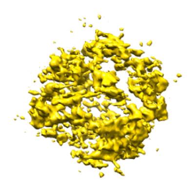
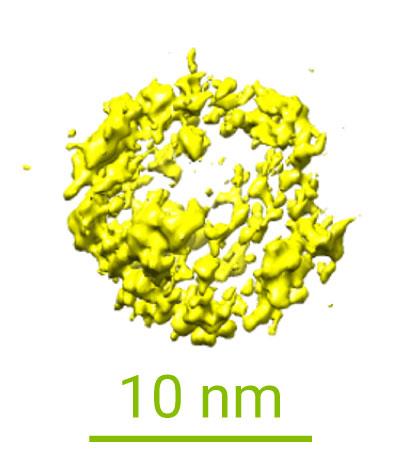
Proteins play a pivotal role in our physiological conditions and associated diseases. A deeper understanding of the kinetics governing the mechanistic behaviour of proteins in liquid media can lead to big improvements in drug design and ultimately in general healthcare.

*This manuscript is currently being updated with long molecular dynamics simulations of ferritin in solution.

The new Stream system

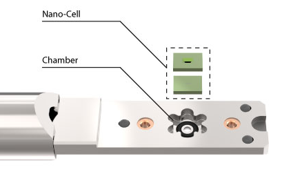
Now the group is advancing to the DENSsolutions Stream system, allowing them to do new kinds of experiments. The big advantage of the Stream system is that it can control the bulging of the viewing windows and therefore the liquid thickness. Controlling the bulging is essential for creating reproducible results. In previous LPEM in situ systems, the window bulging could differ between experiments, thus preventing experiment reproducibility.

Now with the Stream system, the bulging can be adjusted precisely for each new experiment, guaranteeing the same level of bulging and, therefore, consistent results. Controlling the liquid thickness is also important to achieve high contrast in organic and biological materials. The liquid thickness can be reduced up to the equilibrium where you have the highest possible resolution combined with a thick enough layer to have a realistic sample environment. 

Learn more about our Stream system:

Discover publications in which our Ocean system was used:

Do you want to receive interesting articles like this in your mailbox? Subscribe to our newsletter.

Membranes made from Nano-droplets have potential in Medical Research

Membranes made from Nano-droplets have potential in Medical Research

Membranes formed in-lab from nano-droplets could have future use in medicines

For the first time, researchers from the Laboratory of Materials and Interface Chemistry, Eindhoven University of Technology (TUE), with a significant contribution of assistant professor Joe Patterson, have made a comprehensive video of liquid membrane formation using a transmission electron microscope (TEM). They used soap-like nanodroplets submerged in water to create the membrane. Their results are published in Nature, Chemistry and have been highlighted in the Nature, Chemistry News & Views article ‘The molecular Lego movie’.

LPEM Movie of the in-situ self-assembly experiment. Stabilized and cropped. Ianiro, A. et al. Nat. Chem. (2019)

This experiment has continuously recorded the whole process of how the membrane is formed under a microscope. Before this, scientists had to freeze the final membrane and get a snapshot of one or several moments of the membrane forming. This advance is achieved due to a well controlled liquid environment and can be now set in the microscope thanks to the DENSsolutions Ocean system.

Screenshots from the video of the membrane forming on the silicon chip. These were taken using a transmission electron microscope. You can watch the full video here. Ianiro, A. et al. Nat. Chem. (2019)
Membranes are of great interest in research as their selective barriers have potential uses in many fields: drug delivery, water treatment and chemical processes all rely on membrane technology. They are of particular interest in pharmaceutical research as they are the ideal shape to transport a drug through the body and release it only when the membrane finds a specific type of cell, for example, a cancer cell.

The Experiment

The researchers from the Materials and Interface Chemistry group led by Prof. Nico Sommerdijk formed a membrane from soap-like molecules called amphiphilic molecules, which simply means that they interact with both fats and water. Amphiphilic molecules are good building blocks for membranes as they can be lined up with the water-interacting side facing one way and the lipid-interacting parts facing the other way to form larger structures.

The DENSsolutions Ocean In Situ TEM liquid system was essential in this research. The core of the system consists of a dual chip Nano-Cell that sandwiches two chips together to form a microfluidic compartment.

First, the chambers within the tip surrounding the Nano-Cell were flooded with an amphiphilic solvent in order for it to fill the compartment. Then, the solvent was expelled with air, leaving the compartment saturated. Finally, the tip was flooded with water which gradually diffused into the compartment. It was during this stage that the water particles encouraged the solvent particles to organise themselves into a membrane structure.

Step 1. Polymer solvent

Step 2. Air

Step 3. Water

The membrane itself formed in stages. First, the solvent molecules arranged themselves into nanodroplets with a hydrophobic core and a protective hydrophilic shell. The DENSsolutions Nano-Cell created a hotspot of these nanodroplets and they gradually arranged themselves into a hollow membrane.
Diagram of the amphiphilic membrane forming in water. Arash Nikoubashman and Friederike Schmid.

Future Research

Watching how the nanoparticles form and arrange themselves with an electron microscope is a huge step in learning how to manipulate these membranes. The techniques covered in this research will be of interest to scientists working in food science, synthesis chemistry and separation science.

Hanglong Wu, who made a significant contribution to this paper during his PhD period, commented in an interview with DENSsolutions, that the technique “has been extensively used in studying the dynamics and structures of hard materials (for example, metallic nanoparticles) in the aqueous solution in the last decade, but it has been barely employed into soft matter field, mainly due to the inherent high beam sensitivity and low contrast.

“In this Nat. Chem. paper, we actually demonstrate we can probe the soft matter formation with such high contrast. People for sure will start to use the technique in the soft matter field.” – Hanglong Wu

The next stage will be fine-tuning how to manipulate the size and shape of the membrane. This research from Eindhoven is an important step in an exciting field.

If you are interested in the equipment we provided for this research, then contact us to see how we can streamline your experiments.

Membranes formed in-lab from nano-droplets could have future use in medicines

For the first time, researchers from the Laboratory of Materials and Interface Chemistry, Eindhoven University of Technology (TUE), with a significant contribution of assistant professor Joe Patterson, have made a comprehensive video of liquid membrane formation using a transmission electron microscope (TEM). They used soap-like nanodroplets submerged in water to create the membrane. Their results are published in Nature, Chemistry and have been highlighted in the Nature, Chemistry News & Views article ‘The molecular Lego movie’.

LPEM Movie of the in-situ self-assembly experiment. Stabilized and cropped. Ianiro, A. et al. Nat. Chem. (2019)

This experiment has continuously recorded the whole process of how the membrane is formed under a microscope. Before this, scientists had to freeze the final membrane and get a snapshot of one or several moments of the membrane forming. This advance is achieved due to a well controlled liquid environment and can be now set in the microscope thanks to the DENSsolutions Ocean system.

Screenshots from the video of the membrane forming on the silicon chip. These were taken using a transmission electron microscope. You can watch the full video here. Ianiro, A. et al. Nat. Chem. (2019)
Membranes are of great interest in research as their selective barriers have potential uses in many fields: drug delivery, water treatment and chemical processes all rely on membrane technology. They are of particular interest in pharmaceutical research as they are the ideal shape to transport a drug through the body and release it only when the membrane finds a specific type of cell, for example, a cancer cell.

The Experiment

The researchers from the Materials and Interface Chemistry group led by Prof. Nico Sommerdijk formed a membrane from soap-like molecules called amphiphilic molecules, which simply means that they interact with both fats and water. Amphiphilic molecules are good building blocks for membranes as they can be lined up with the water-interacting side facing one way and the lipid-interacting parts facing the other way to form larger structures.

The DENSsolutions Ocean In Situ TEM liquid system was essential in this research. The core of the system consists of a dual chip Nano-Cell that sandwiches two chips together to form a microfluidic compartment.

First, the chambers within the tip surrounding the Nano-Cell were flooded with an amphiphilic solvent in order for it to fill the compartment. The solvent was then expelled with air, leaving the compartment saturated. Then, the solvent was expelled with air, leaving the compartment saturated. Finally, the tip was flooded with water which gradually diffused into the compartment. It was during this stage that the water particles encouraged the solvent particles to organise themselves into a membrane structure.

Step 1. Polymer solvent

Step 2. Air

Step 3. Water

The membrane itself formed in stages. First, the solvent molecules arranged themselves into nanodroplets with a hydrophobic core and a protective hydrophilic shell. The DENSsolutions Nano-Cell created a hotspot of these nanodroplets and they gradually arranged themselves into a hollow membrane.
Diagram of the amphiphilic membrane forming in water. Arash Nikoubashman and Friederike Schmid.

Future Research

Watching how the nanoparticles form and arrange themselves with an electron microscope is a huge step in learning how to manipulate these membranes. The techniques covered in this research will be of interest to scientists working in food science, synthesis chemistry and separation science.

Hanglong Wu, who made a significant contribution to this paper during his PhD period, commented in an interview with DENSsolutions, that the technique “has been extensively used in studying the dynamics and structures of hard materials (for example, metallic nanoparticles) in the aqueous solution in the last decade, but it has been barely employed into soft matter field, mainly due to the inherent high beam sensitivity and low contrast.

“In this Nat. Chem. paper, we actually demonstrate we can probe the soft matter formation with such high contrast. People for sure will start to use the technique in the soft matter field.” – Hanglong Wu

The next stage will be fine-tuning how to manipulate the size and shape of the membrane. This research from Eindhoven is an important step in an exciting field.

If you are interested in the equipment we provided for this research, then contact us to see how we can streamline your experiments.

239